Congratulations to Ewa who got a Preludium grant in the last round to study “Genesis of dendritic spine head protrusion (SHP) formation”. In the project Ewa will be studying the influence of glutamate on the formation of spine head protrusions using a novel noninvasive electrochemical glutamate biosensor and fluorescent confocal microscopy methods. The study of SHPs will be done at the Nencki Institute for Experimental Biology, which is also the institute where the project is formally located. In our lab she will develop a biosensor for measuring glutamate levels locally in neurocell cultures.
The popular science version of the project can be read below.
Genesis of dendritic spine head protrusion (SHP) formation
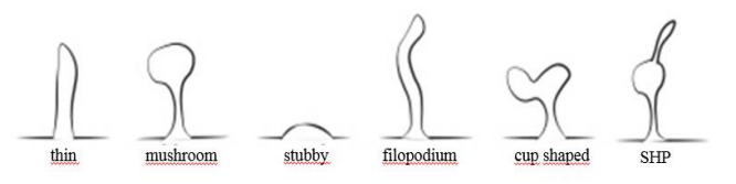
One of the most important functions of the nervous system is the ability to receive, process and store information provided from the external environment. Communication of nerve cells is possible thanks to specialized connections called synapses. Synaptic plasticity underlies the brains ability to learn and remember, and is understood as the ability of nerve cells to modify the strength of synaptic connections. Most excitatory synapses in the mammalian brain are located on the dendritic spines, which are their postsynaptic part. In the structure of dendritic spines we can distinguish head and neck which binds the head with the surface of dendrites. The shape and number of dendritic spines reflects the strength of synaptic connections. One of the morphological forms of dendritic spines is the filopodium on the top of the spine head, so-called spine head protrusion (SHP) (Figure 1.). Our research shows that SHP occurs as a result of neuronal stimulation and that SHP is capable of creating new synaptic connections, independently of those already existing on a given dendritic spine. The role of SHPs in synaptic plasticity and the mechanisms regulating their formation have not been unambiguously described so far. However, it is speculated that glutamate, main excitatory neurotransmitter in the brain, may play a crucial role in their formation processes.
In our project, we will determine the genesis of SHP formation in relation to glutamate concentration. For this means we will develop a electrochemical tool enabling detection of local, extracellular glutamate concentration in primary neuronal cultures. Electrochemical methods allow to obtain real-time information, often with high spatial and temporal resolution and can serve as an excellent supplement to the traditional optical analysis used for in vitro studies. In our project, we would like to develop electrophysiologically non-invasive glutamate biosensor for short-term glutamate measurements. The proposed system will allow spatial positioning of the biosensor probe under live-cell imaging confocal microscopy conditions simultaneously enabling observation of structural changes in dendritic spines, including SHP formation and quantification of glutamate.
Glutamate plays a key role in synaptic plasticity. Depending on the concentration, it may regulate both physiological and pathological processes (learning and memory, neurodegeneration). The minimally invasive method developed by us gives the opportunity to accurately determine glutamate concentrations within individual synapses in neuronal cultures in vitro, thus being an excellent tool for testing synaptic plasticity. The unique properties of the solution proposed by us will allow to determine the genesis of SHP, which will directly contribute to a better understanding of the mechanisms responsible for the creation of new synaptic connections underlying the proper functioning of the nervous system.

