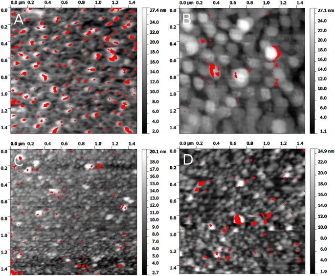
A paper was recently published in Scientific Reports about atomic force microscopy, investigating interactions between bacteriophages and bacteria. Or, at least, as the title says, between T4 adhesin with lipopolysaccharides from bacteria. This was done by modifying an AFM tip with the adhesin from the bacteriophage T4 and using this tip to scan a sample surface modified with lipopolysaccharides. The interaction between the surface and the tip as well as the topography was measured simultaneously using the PicoTREC AFM mode. The force needed to pull apart the adhesin and the target was measured using force spectroscopy. By combining these to techniques we could see that the points of high interaction was correlated with the positions of the polysaccarides, and we could measure this interaction. The forces between the adhesin from T4 and a lipopolysaccharide from one bacterium specifically target by T4 and two non-specific bacteria were measured, and the result shows that a higher force is needed to separate the specifically interacting pair.
In this paper, most of the work was done by Ewa Brzozowska from Wrocław and Adam Leśniewski from the Nanosurface engineering group at our institute. Martin had just a small part in the paper, mainly analysing the data.